Improvement of skin texture and wrinkles using radiofrequency ultra-thin electrode technology
Abstract
Background: Application of radiofrequency (RF) skin tightening energy with microneedles to facial skin shown to promote skin renewal.
Aim: To evaluate the safety and efficacy of the VoluDerm technology with 100 ultra-thin electrodes gen100 tip for improvement of skin texture and wrinkles. Methods: A prospective, open-label, intra-individual-controlled trial. Twelve subjects (mean age, 45.5; Fitzpatrick skin types II-III) with Fitzpatrick Elastosis Scale (FES) score 3-6 were treated with VoluDerm RF technology using the gen100 ultra-thin electrodes disposable tip. The participants underwent three treatments at 3-week intervals. Treatment safety was evaluated at each treatment visit and at the follow-up visits (1 and 3 months post last treatment). Efficacy of treatment evaluated at 1 and 3 months after the last treatment using the FES as well as evaluator- and subject-rated Global Aesthetic Improvement Scale (GAIS).
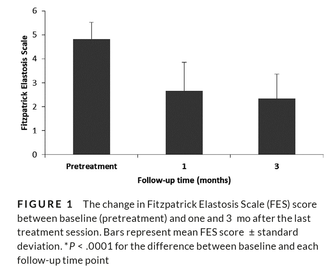
Results: Treatment was well tolerated, with no downtime or adverse events. One and three months after the last treatment session, physician-rated FES showed statisti-cally significant improvement of 2.67 ± 1.18 and 2.33 ± 1.03, respectively (P < .0001), from baseline (4.83 ± 0.69). According to the investigator assessment of improve-ment using the GAIS scale, all subjects had an improvement in skin texture and pig-mentation and most of the patients (91.7%) had an improvement in skin brightness, tightness, and wrinkles.
Conclusion: This is the first study evaluating the Legend+ RF System with VoluDerm technology using the novel gen100 ultra-thin electrodes tip. This study demonstrated effective and well-tolerated treatment for improving skin texture and appearance with minimal to no downtime.
K E Y WO R D S
facial rejuvenation, microneedling device, resurfacing, RF, skin texture, ultra-thin electrodes, eye wrinkle reduction device
INTRODUCTION
The growth in the aging population together with an increased life span has brought about a growing demand for rejuvenation treat-ments. A wide range of noninvasive and nonsurgical technologies are currently being developed and used for skin rejuvenation in order to improve the appearance of the skin, particularly facial skin. Many of these technologies are based on processes that cause skin resurfacing and dermal collagen remodeling. Ablative resurfacing lasers, such as carbon dioxide and erbium: yttrium aluminum gar-net lasers, completely remove the skin’s surface in a controlled manner,1,2 however require prolonged recovery, and can lead to an increased risk of depigmentation, scarring, and infections.3 To decrease such adverse effects, newer treatment modalities have emerged.
Fractional radiofrequency (RF) technologies use an array of mi-croneedles that penetrate the skin and heat the deep dermis, cre-ating zones of invisible thermal microwounds, characterized by an optimized dermal impact and minimal epidermal effect, between areas of unaffected normal healthy skin. These healthy skin zones function as healing centers, promoting neocollagenesis and skin re-newal in a safe and effective manner.4-6 This technology has demon-strated efficacy when used for skin resurfacing, reducing wrinkles and enhancing skin and facial appearance.7
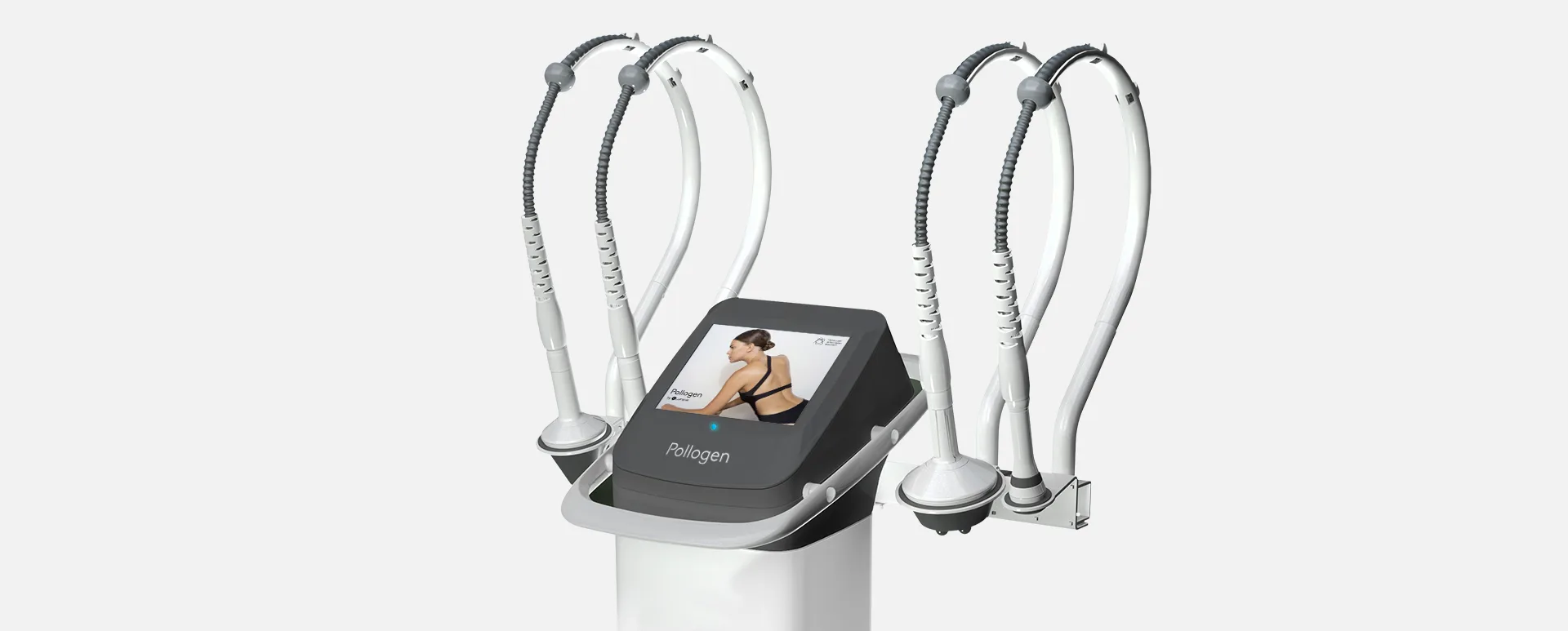
The unique Legend+ VoluDerm™ technology (Pollogen Ltd), aimed for dermatological procedures requiring coagulation, ablation, and resurfacing of the skin, uses disposable tips that are attached to the distal end of the applicator and placed on the target skin area. The tip comprises a high-density array of ultra-thin electrodes (150 µm width and different lengths) that smoothly penetrate the skin via RF-assisted ablation, instead of the traditional mechanical force used with other technologies. The ultra-thin electrodes ablate pinpoint-sized spots in the dermal layer, inducing coagulation and stimulating the natural wound healing process and the production of hyaluronic acid, new collagen, and elastin. This process results in significant dermal volumization, wrinkle reduction, and skin rejuvenation.
The effect of VoluDerm RF ultra-thin electrodes technology for treating skin texture, laxity, and wrinkle reduction was previously demonstrated with gen 36 disposable tip, composed of an array of 36 ultra-thin electrodes and (0.6 mm in length and 150 µm width). The treatment was safe and effective for dermal volumizing and treatment of wrinkles with minimal pain and no downtime.8
This is the first study to evaluate the safety and efficacy of the VoluDerm applicator with the gen100 disposable tip, a high-density array of 100 ultra-thin electrodes for improvement of skin texture and wrinkles.
2 | MATERIAL S AND METHODS
2.1 | Design and subjects
This was a single-center, prospective, open-label, intra-individual-controlled trial, approved by the Institutional Review Board (IRB). All of the participants signed an informed consent form prior to receiv-ing the treatments.
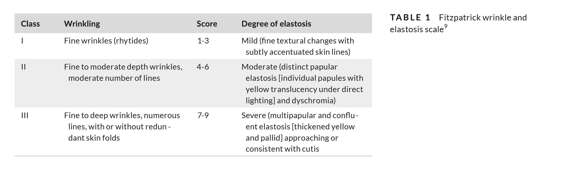
Inclusion criteria for the study included healthy adult subjects, 30-50 years of age with Fitzpatrick skin types I-VI, with facial wrinkles, and Fitzpatrick Elastosis Scale (FES)9 score 3-6 (Table 1). Exclusion criteria included pregnancy or breast-feeding, any im-plantable electronic device, or any active dermatological disorder in the treatment site.
2.2 | Treatment protocol and settings
The entire face was treated, including the perioral and periorbital regions. The subjects received three treatments, at 3-week inter-vals, with follow-up at 1 and 3 months after the last treatment. Prior to each treatment, the treatment area was cleaned and dried. Use of a topical anesthetic agent (benzocaine 20%, lidocaine 6%, or tet-racaine 4%) was allowed according to patients’ request.
Treatment settings: During the first treatment, a single pass over the treatment area (with no overlap) was performed using the sys-tem’s low-setting preset parameters. During the second treatment, a single pass with 50% overlap was done, using the system’s low-set-ting preset parameters for the periorbital region, and medium-set-ting preset parameters for the perioral region and other facial areas. During the third treatment, two passes over the treatment area were performed using low-setting preset parameters for the periorbital region, medium-setting preset parameters for the perioral region, and elevated medium setting (30% power and 30% exposure) for rest of the facial areas.
After each treatment, the subjects were instructed to avoid makeup and moisturizer for about 24 hours. The subjects were in-structed to cover the treated skin with a high-factor sunscreen, after 24 hours.
2.3 | Study endpoints and outcome measures
Safety endpoint included the evaluation of immediate skin response and subjects’ assessment of pain and discomfort associated with the treatments, using a Pain Visual Analogue Scale (VAS), where 0 is “no pain” and 10 is “intolerable pain.” Skin safety throughout the study
See also: Oxygen Therapy Treatment Machines (Oxygeneo)
was evaluated by examining the posttreatment occurrences of com-plications and adverse events.
The efficacy primary endpoint of the study was the success rate at the 3-month follow-up visit, with success defined as an im-provement of at least 1 point on the FES assessed by investigator as compared to baseline. The secondary endpoint of the study was skin improvement on the FES at the 1-month follow-up visit as compared to baseline.
The subjects were photographed at each treatment visit and at the 3-month and 1-month follow-up visits.
Two blinded uninvolved evaluators evaluated each subject’s improvement in skin texture and wrinkles by comparing the pho-tograph taken at the follow-up visits to the photograph taken at baseline.
Evaluators rated the changes in skin texture and wrinkles using the Fitzpatrick Elastosis Scale (FES; Table 1).
The GAIS scale was used independently by the investigator and subjects for grading the changes in skin appearance (0 = worse, 1 = no change, 2 = improved, 3 = much improved, 4 = very much improved).
Treatments’ downtime was defined as the period of time follow-ing the procedure during which the subject had edema and erythema and felt unable or unwilling to go out in public.
2.4 | Statistical analysis
Statistical analyses were mainly descriptive in nature, with continu-ous variables summarized by mean and standard deviation, and cat-egorical variables by count and percentage. The difference between baseline and follow-up was analyzed using t test. The level of signifi-cance was 5%.
3 | RESULTS
Twelve Caucasian females age 39-58 (mean age 45.5 ± 5.2) and Fitzpatrick skin types II-III were included in the study. Baseline phy-sician-assessed FES was 4.83 ± 0.69. None of the subjects received pretreatment anesthesia prior to the first treatment, one subject (8.3%) received pretreatment anesthesia prior to the second treat-ment, and five subjects (41.7%) received pretreatment anesthesia prior to the third treatment.
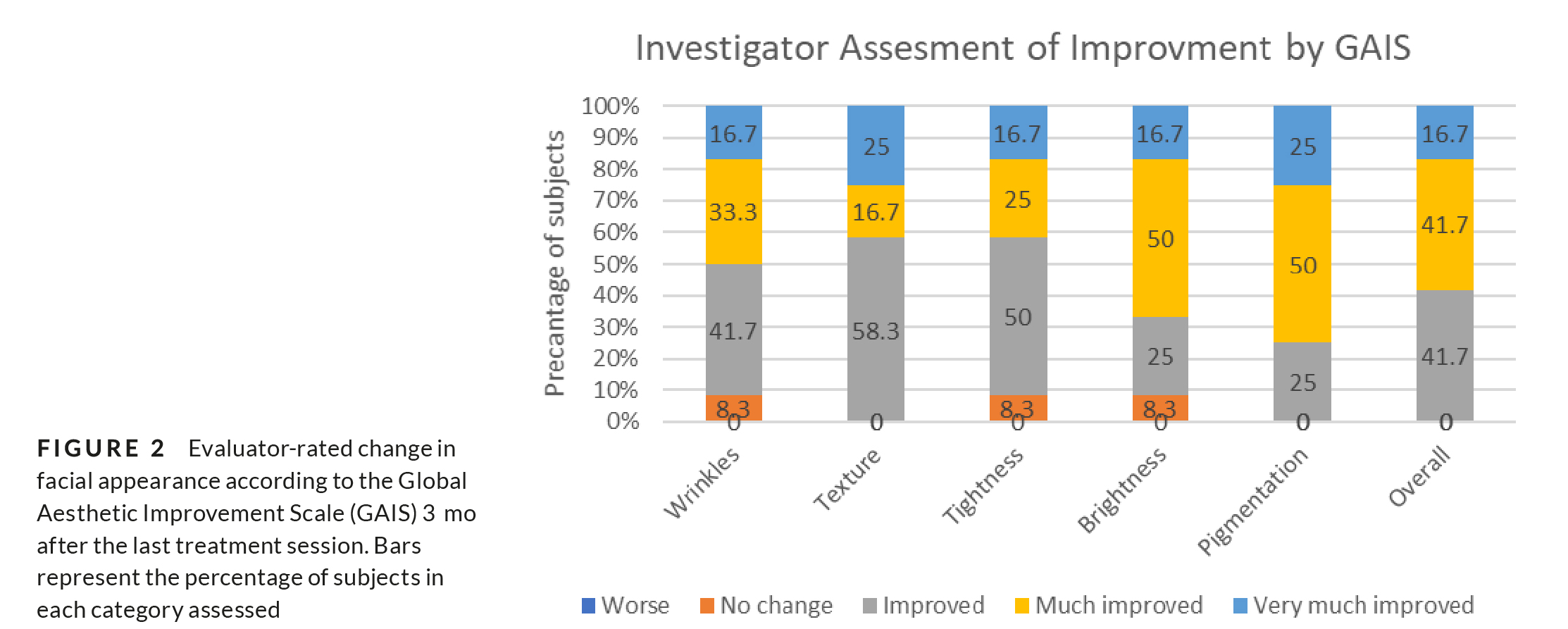
One and three months after the last treatment session, mean physician-rated FES was 2.67 ± 1.18 and 2.33 ± 1.03, respectively (P < .0001; Figure 1). According to investigator assessment of im-provement by GAIS scale at the 3-month follow-up, skin texture was improved in 58.3% of the subjects, much improved in 16.7%, and very much improved in 25%. Skin pigmentation was improved in 25% of the subjects, much improved in 50%, and very much improved in 25%. Skin tightness, brightness, and wrinkles were improved in 92.7% of patients, and overall improvement was observed in all pa-tients in various degrees (Figure 2).
Patient’s satisfaction rate from the treatment was high. Six of the subjects (50%) reported an improvement in skin texture already after one treatment and 83.3% reported such improvement after two treatments.
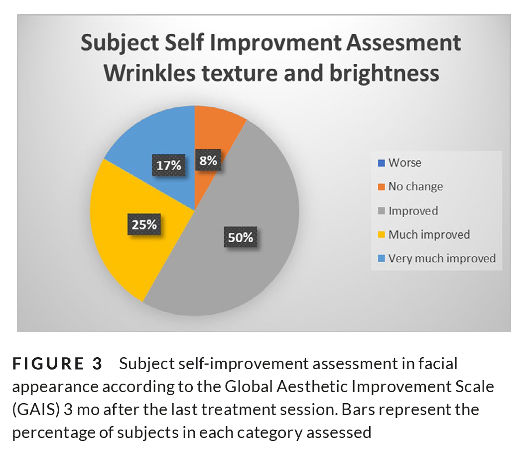
According to subject self-improvement GAIS at the 3-month follow-up, skin texture and wrinkles were very much improved in 16.7% of the subjects, much improved in 50%, improved in 16.7%, whereas only 8.3% reported on no change (Figure 3).
Blinded evaluation of the baseline photographs compared with the 3-month follow-up photographs showed a mean reduction of 1.63 points in FES from baseline.
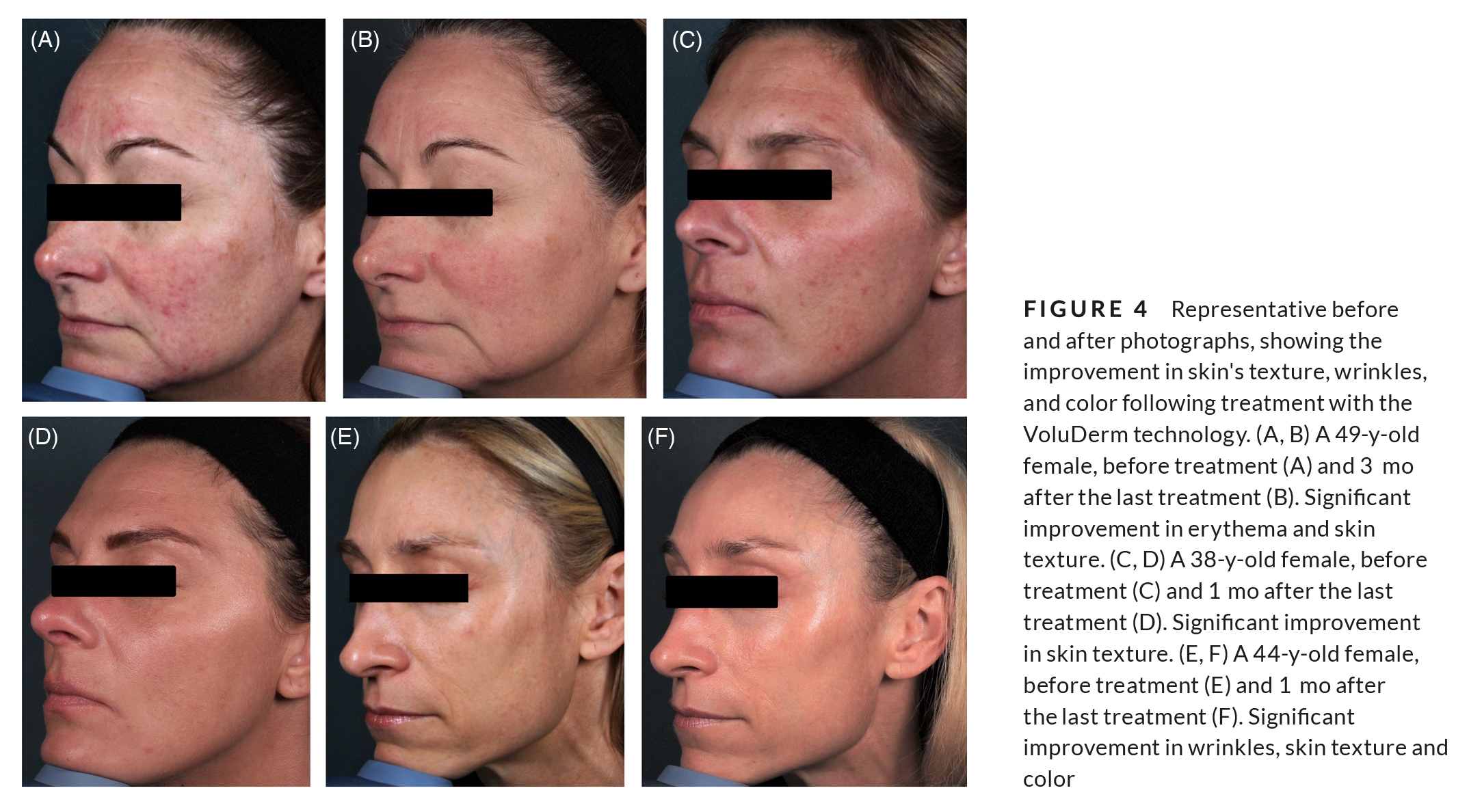
The before and after photographs demonstrate improvement in skin texture, color, and wrinkles (Figure 4).
3.1 | Safety
No other adverse events were reported. No downtime was reported for any of the subjects, and all of them felt comfortable to go out in public. Mean pain was greater during the third treatment as com-pared to the first treatment and was highest in the perioral region followed by the periorbital region (Table 2).
4 | DISCUSSION
This study has shown that three consecutive treatment sessions at 3-week intervals using VoluDerm technology for skin applicator with gen100 ultra-thin electrodes significantly improve skin texture, wrinkles, brightness, and pigmentation. Improvement of skin parameters started from the first treatment and lasted for at least 3 months after the last treat-ment session (the last time point assessed in the study). Moreover, treatment was well-tolerated, with no downtime nor adverse events. Pain levels during treatment slightly increased with each treatment session, which can be correlated with the increase in treatment pa-rameters and number of passes and overlaps. Nevertheless, pain lev-els remained low throughout the study.
Studies in animal models have shown that RF microneedling triggers the body’s natural healing response involving inflammatory, proliferative, and remodeling stages.10 Complete healing, namely subepidermal healing with epidermal and dermal regeneration, was demonstrated within 14 days for various energy levels. The extent of effect showed a correlation with the energy level used.10 Collagen re-modeling was induced with an increase in mid-to-deep dermis glycos-aminoglycans, as well as epidermal mitotic index.11 RF microneedles
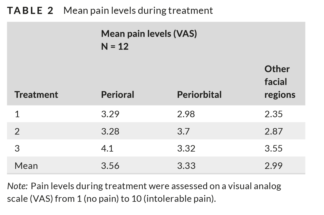
Note: Pain levels during treatment were assessed on a visual analog scale (VAS) from 1 (no pain) to 10 (intolerable pain).
caused immediate (Day 0) epidermal and dermal effects, followed by a progressive healing process. Dermal RF-induced coagulated columns showed mixed cellular infiltration, neovascularization, and granula-tion tissue formation.12 Another study reported increased epidermal hyaluronan and CD44 expression in both young and old aged mice, suggesting that these factors have a role in overcoming age-related epidermal dysfunction by recovering barrier homeostasis.13 Clinical studies employing minimally invasive RF microneedling have shown success in treatment of wrinkles and skin laxity. The number of needles used varies between 10-64 with needles-length as long as 3.5 mm.4,5,14-18
The present study has shown for the first time skin-tightening fol-lowing the use of VoluDerm applicator with gen100 disposable tips, a high-density array of 100 ultra-thin electrodes. The VoluDerm RF-assisted ultra-thin electrodes technology mainly affects the dermal layer,10 while the epidermal barrier functions are only marginally af-fected; thus, the treatment is associated with little to no downtime, reduced discomfort, and less predisposition to side effects compared to fractional and RF microneedling traditional technologies. While with traditional microneedling the penetration mechanism is mechan-ical, with the VoluDerm technology the RF-assisted ultra-thin elec-trodes ablate the epidermal layer all through to the deep dermis. Both evaluator-rated and subject-rated results clearly and unequivocally indicated that treatment with VoluDerm applicator and gen100 dis-posable tip, high-density RF-assisted ultra-thin electrodes, is well-tol-erated and effective in improving skin texture and appearance. Longer assessment of the treatment’s effect duration is warranted.
ACKNOWLEDGMENTS
Dr Gold is a consultant and performed clinical research for Pollogen/Lumenis for this project.
ORCID
Michael H. Gold
REFERENCES
1. Nanni CA, Alster TS. Complications of carbon dioxide laser resurfacing. An evaluation of 500 patients. Dermatol Surg. 1998;24(3):315-320.
2. Tanzi EL, Alster TS. Side effects and complications of variable-pulsed erbium:yttrium-aluminum-garnet laser skin resurfacing: extended experience with 50 patients. Plast Reconstr Surg. 2003;111(4):1524-1529; discussion 1530-1532.
3. Metelitsa AI, Alster TS. Fractionated laser skin resurfacing treat-ment complications: a review. Dermatol Surg. 2010;36(3):299-306.
4. Hantash BM, Ubeid AA, Chang H, Kafi R, Renton B. Bipolar frac-tional radiofrequency treatment induces neoelastogenesis and neocollagenesis. Lasers Surg Med. 2009;41(1):1-9.
5. Hantash BM, Renton B, Berkowitz RL, Stridde BC, Newman J. Pilot clinical study of a novel minimally invasive bipolar microneedle ra-diofrequency device. Lasers Surg Med. 2009;41(2):87-95.
6. Hruza G, Taub AF, Collier SL, Mulholland SR. Skin rejuvenation and wrinkle reduction using a fractional radiofrequency system. J Drugs Dermatol. 2009;8(3):259-265.
7. Gold MH, Adelglass J. Evaluation of safety and efficacy of the TriFractional RF technology for treatment of facial wrinkles. J Cosmet Laser Ther. 2014;16(1):2-7.
8. Shapiro S. VoluDerm micro-needle technology for treating skin laxity and wrinkles-initial clinical experience. Jacobs Journal of. Exp Dermatol. 2015;1(2):1-7.
9. Fitzpatrick RE, Goldman MP, Satur NM, Tope WD. Pulsed carbon dioxide laser resurfacing of photo-aged facial skin. Arch Dermatol. 1996;132(4):395-402.
10. Gershonowitz A, Gat A. VoluDerm microneedle technology for skin treatments-in vivo histological evidence. J Cosmet Laser Ther. 2015;17(1):9-14.
11. Boisnic S, Branchet MC. Ex-vivo study of hybrid energy technology using a human skin model. Eur J Dermatol. 2014;24(1):46-52.
12. Zheng Z, Goo B, Kim DY, Kang JS, Cho SB. Histometric analysis of skin-radiofrequency interaction using a fractionated microneedle delivery system. Dermatol Surg. 2014;40(2):134-141.
13. Lee HJ, Seo SR, Yoon MS, Song JY, Lee EY, Lee SE. Microneedle fractional radiofrequency increases epidermal hyaluronan and reverses age-related epidermal dysfunction. Lasers Surg Med. 2016;48(2):140-149.
14. Seo KY, Yoon MS, Kim DH, Lee HJ. Skin rejuvenation by micronee-dle fractional radiofrequency treatment in Asian skin; clinical and histological analysis. Lasers Surg Med. 2012;44(8):631-636.
15. Seo KY, Kim DH, Lee SE, Yoon MS, Lee HJ. Skin rejuvenation by microneedle fractional radiofrequency and a human stem cell con-ditioned medium in Asian skin: a randomized controlled investigator blinded split-face study. J Cosmet Laser Ther. 2013;15(1):25-33.
16. Lee HS, Lee DH, Won CH, et al. Fractional rejuvenation using a novel bipolar radiofrequency system in Asian skin. Dermatol Surg. 2011;37(11):1611-1619.
17. Gold M, Taylor M, Rothaus K, Tanaka Y. Non-insulated smooth mo-tion, micro-needles RF fractional treatment for wrinkle reduction and lifting of the lower face: international study. Lasers Surg Med. 2016;48(8):727-733. 18. Tanaka Y. Long-term three-dimensional volumetric assessment of skin tightening using a sharply tapered non-insulated microneedle radiofrequency applicator with novel fractionated pulse mode in Asians. Lasers Surg Med. 2015;47(8):626-633.